Describe the Structure of the Small Intestine
- Aggregation of lymphoid tissue. The small intestine is divided into the duodenum jejunum and ileum.

Small Intestine Definition Examples Diagrams
Distinctive features of the ileum.

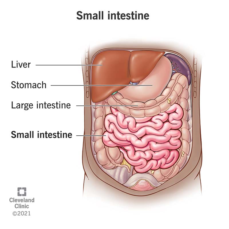
. In living humans the small. Made up of three segments the duodenum jejunum and ileum the small intestine is a 22-foot long muscular tube that breaks down food using enzymes released by the pancreas and bile from the liver. All three parts are covered with the greater omentum anteriorly.
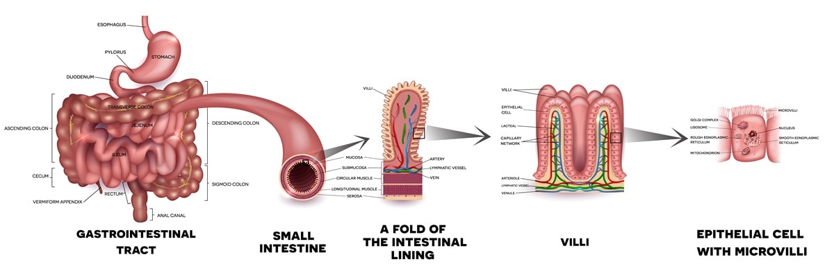
The inner surface of the small intestine is not flat but thrown into circular folds which not only increase surface area but aid in mixing the ingesta by acting as baffles. The small intestine is made up of the duodenum jejunum and ileum. The small intestine is divided into three parts.
Together with the esophagus large intestine and the stomach it forms the gastrointestinal tract. The wall of the small intestine is composed of the same four layers typically present in the. The Small Intestine Structure.
The duodenum has both intraperitoneal and retroperitoneal parts while the jejunum and ileum are entirely intraperitoneal organs. Finger-shaped structures called villi line the entire small intestine. There are specialized structures in small intestine that makes it better adapted for absorption.
The inner surface is thrown into a series of circular folds. The large intestine however lacks these structures. Small Intestine Large Intestine Bladder The permeability of epithelia varies between organs up to 10000 fold dierence.
Distinctive features of the jejunum. Much of the small intestine is covered in projections called villi that increase the surface area of the tissue available to absorb nutrients from the gut contents. The small intestine has small tiny projections called villi.
However studies of its biology are limited by the lack of a feasible method to visualize all the relevant components for its regulation. Submucosa Connective tissue layer which contains blood vessels lymphatics and the submucosal plexus. - Has the tallest villi.
From proximal at the stomach to. Structure of small intestine. Peristalsis also works in this organ moving food through and mixing it with digestive juices from the pancreas and liver.
- Increased number of goblet cells. 1 Villi and microvil View the full answer. The small intestine is the longest part of the digestive system measuring about 20 ft.
Answer Small intestine is the part of digestive tract involved in digestion and absorption of all the macronutrients. There are 24 dierent claudin genes and each encodes a protein with dierent permeability properties. The epithelia of the small intestine is more permeable that the epithelia of the bladder.
Structure of the small intestine. Here we describe an efficient whole-mount protocol to visualize the intact structure of lacteals and surrounding cells in. The small intestine is part of the digestive system and is mainly responsible for the absorption of nutrients.
This is where the final parts of digestive absorption take place. Up to 25 cash back The small intestine incorporates three features which account for its huge absorptive surface area. The ileum absorbs bile acids fluid and vitamin B-12.
Folds villi and micro villi. The mucosa forms multitudes of projections. There are villi and microvilli that line the small intestine.
Learn about the structure of. The coiled tube of the small intestine is subdivided into three regions. Some claudins are selective for.
Internal structure of the small intestine. The duodenum is the first segment. Longest part of the GI tract first part duodenum second part jejunum third part ileum Functions of small intestine.
- Very few lymphoid follicles. The small intestine is the section of your digestive tract where the majority of food digestion and nutrient absorption takes place. The outer longitudinal layer and inner.
- Lots of Peyers patches. The lowest part of your small intestine is the ileum. The small intestine is divided into the duodenum jejunum and ileum.
- There are no Brunner glands. State how the absorptive surface area of the small intestine is increased. Learn vocabulary terms and more with flashcards games and other study tools.
Muscularis externa Consists of two smooth muscle layers. This is for increased surface area for the maximum aborbsion by the small intestine. Describe the structure of the interior surface of the small intestine and explain how this structure relates to its function.
These projections increase the surface area for absorption. The small intestine is a windingtightly folded tube about 20ft6m long in adultsIt connects to the stomach on the top end and to the large intestine colon on the bottom endMost of the food a person consumes is digested and absorbed in the small. Mucosa Innermost layer Contains the epithelium lamina propria and muscularis mucosae.
Villi are units of absorption of food which are present in the small intestine. Start studying Structure and function of the Small intestine. Together these can extend up to six meters in length.
Absorbs products of digestion chemical digestion absorption of proteins lipids and carbohydrates. It is present in the central and lower abdominal cavity.

What Does The Small Intestine Do

Small Intestine Function Anatomy Definition
Small Intestine Structure Function Digestive System Anatomy And Physiology
0 Response to "Describe the Structure of the Small Intestine"
Post a Comment